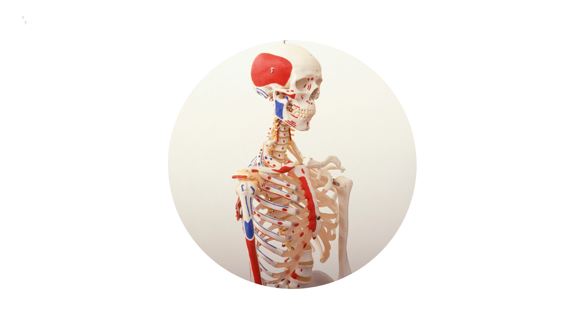
THE SKELETON
The skeleton is made up of 206 bones joined together by joints, it’s the body armor, gives it its shape and determines the height of the individual.
The functions of the skeleton are:
- Support and shape the body.
- Protect internal organs such as the brain, lungs, heart…
- Biomechanical base of movement support: anchoring to the muscles, they pull the bones to which they are attached and we can perform all kinds of movements.
- Hematopoiesis: formation of blood cells.
- Mineral reserve of the organism.
THE BONE CELL AND GROWTH PERIODS
The bone cell is made up of by:
- 35% by organic substances, mainly by the so-called bone matrix important in traction.
- The remaining 70% is composed of inorganic substances, such as phosphates and mainly important calcium in compression. (45% MINERALS + 20% WATER)
Throughout life, bone formation (ossification) and destruction of the itself (reabsorption) occur simultaneously. The continuous process of destroying the old tissue and creating new one is known as remodeling.
Bone cell types:
- Osteoblasts: Bone-forming cells.
- Osteoclasts: Cells that reabsorb bone.
- Osteocytes: Mature bone cells.
The organism has several phases or periods of growth characterized by:
- A period of fast growth, the firsts four years of life: characterized by a progressive decrease in speed from 25 cm. the first year to 12 cm. the second, 10 cm. the third and 8 cm. the fourth year.
- A period of slower and more sustained growth: from four years to the onset of puberty, with a growth rate that goes between 4.5 – 7.0 cm. /year.
- A new rapid period during pubertal development, in which maximum speed of growth can reach up to 12 cm. /year in the male and 9 cm. /year in the woman.
These opposing processes balance each other from youth to middle age, that is, the rate of bone formation equals the rate of bone destruction itself, therefore, the bones do not grow or shrink, their size remains constant.
With aging there is a degeneration of bone and cartilage, which produces among other things an increased possibility of bone injuries. Walking, running and other kinds of exercise put the bones under stress. They respond by depositing more collagen fibers and mineral salts in the bone matrix.
It has been observed that the number of capillaries that nourish the bones increases if they are under regular effort (training), which could explain the fact that that the injuries of athletes heal much more quickly than those who live a sedentary life.
BONE STRUCTURE
In the bone structure there are two parts:
The compact: which forms a hard and conglomerate mass. It is crossed by a large number of very thin canals, called Haversian canals. These conducts are almost entirely full, by blood vessels, although there are also They may contain marrow tissue or fat. All Haversian canals are communicated with each other, except in the spongy tissue, where there are no such structures.
The spongy: which is arranged in thin columns and plates and is where the red or hematopoietic marrow are located.
Another fundamental element of the bones is the bone marrow, which is a soft mass which occupies the cavity of the bones and the intermediate spaces of the spongy substance of all bones. According to its richness in fat and its function, it is distinguished:
- Yellow marrow: compact, due to its fat content, and mainly occupies the central part of the long bones.
- Red or hematopoietic marrow: more semi-fluid due to its content in precursors cells of the elements that form blood.
TYPES OF BONES
- Long bone: Characterized by its length, appreciating in the same two fundamental parts: the body or diaphysis and the ends or epiphyses. Examples of long bones are the tibia, femur, ulna etc.
- Short Bone: It refers to bones that have more or less cubic being its dimensions (length, width and thickness) sensibly similar. An example of short bones are the calcaneus patella, vertebrae, scaphoid, etc.
- Flat Bone: They are bones whose width and length predominate over thickness. An example of flat bones are the sternum, frontal, shoulder blades, occipital, etc.
All bones are covered by a fibrous tissue called the periosteum. This structure allows muscles to attach to the bones via tendons and It also makes the bone grow thicker.
In long bones we can distinguish the following parts:
- Epiphysis: proximal and distal area of the bone
- Diaphysis: central zone of the bone and formed almost entirely by compact tissue.
- Metaphysis: zone of transition tissue, it is located in the bones in growth, growth plate and in the adult bones without possibility of growth, a transitional tissue in between spongy and compact bone.
HEAD BONES
The head skeleton is the most complex bone structure in the body because surrounds the brain, houses the sensory organs, and surrounds the tracts respiratory digestive orifices.
The skull bones are 8 and constitute a strong box to protect the brain:
- 2 temporary
- Parietal
- Occipital
- Frontal
- Ethmoid
- Sphenoid
The Bones of the face: There are 14 and among them the most important are:
- The jaws (upper and lower) that are used in chewing.
- Lachrymal.
- Nasal, Turbinate and Vomer.
- There is a loose bone at the base of the tongue; called HYOID, in which supported in their movements.
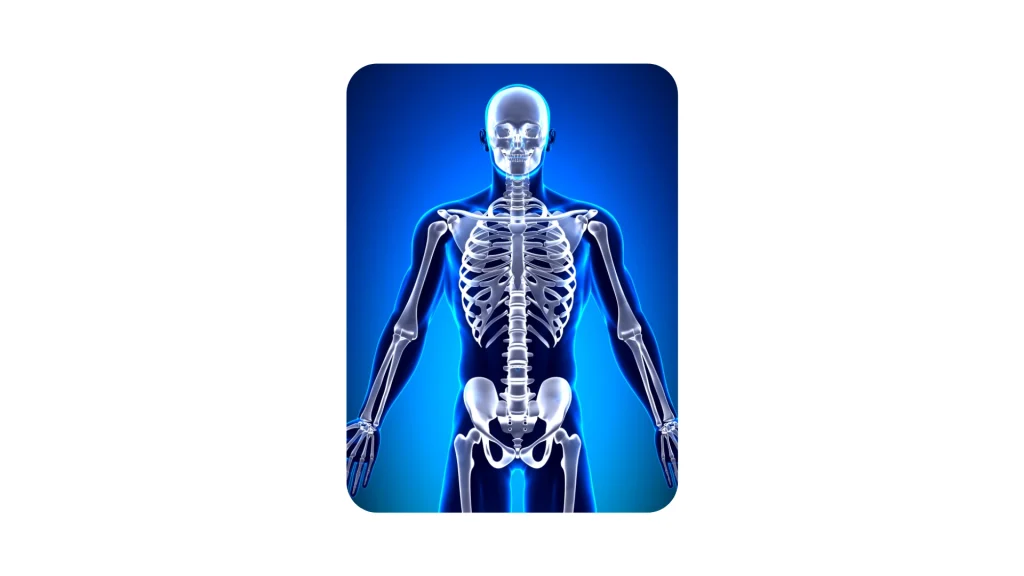
BONES OF THE TRUNK
The clavicle and shoulder blade, which serve to support the upper limbs. The ribs that protect the lungs, forming the rib cage. At the back they will articulate with the thoracic or dorsal vertebrae.
The sternum, where the ribs on both sides meet and protects the structure cardiac (anterior).
RIBS
The ribs are flat bones, very elongated, arch shaped, and it’s back side they articulate with their corresponding dorsal vertebra.
Together with the sternum and the dorsal vertebrae they delimit the thoracic cavity, in which inside, viscera of great importance are housed, such as the heart and lungs.
There are twelve ribs on each side, classified as:
- True ribs, the first seven, articulating with the sternum through the costal cartilages.
- False ribs 8th, 9th and 10th, whose anterior costal cartilage joins the overlying the 6th and 7th ribs
- Floating ribs 11th and 12th, whose anterior costal cartilage remains free and without join the sternum
As a whole each side describes a concave curve inward, and is inclined from top to bottom and back to front. This slope increases progressively from the first to the last rib. Between each of the ribs is a space of about 2 cm. called the intercostal space.
SPINE
The spinal column is made up of a set of juxtaposed bones that called vertebrae, which are articulated and held by muscles and ligaments.
The spine is related to the pelvis below and to the skull above.
It is made up of 33–34 vertebrae that are classified according to their situation, in five regions with their own characteristics in each of them:
- Cervical: Seven vertebrae.
- Dorsal: Twelve vertebrae, articulating in each of them to the rib correspondent.
- Lumbar: Five vertebrae.
- Sacral: Five vertebrae joined together to form one bone.
- Coccygeal: Four-five vertebrae.
The vertebrae are separated from each other by small discs fibrocartilaginous, known as intervertebral discs.
Each vertebra also has a series of articular faces that serve as support and attachment to the upper and lower vertebrae.
Laterally, the junction of each of the vertebrae leaves a conjunction foramen through which the nerve root exits from the spinal cord.
The vertebral column has a vertical direction, but it is not rectilinear, since It presents gentle curvatures in the anterior-posterior direction. The curves directed forward are called lordosis and those directed backwards, kyphosis.
BREASTBONE
It is a flat, elongated bone located in the midline of the anterior chest wall, closing the rib cage in this plane. Its anterior face is convex forward.
UPPER LIMB BONES
- Clavicle, shoulder blade and humerus forming the shoulder joint
- The humerus, in the arm.
- The radius and ulna in the forearm
- The carpus, formed by 8 little bones of the wrist. The metacarpals in the hand.
- Phalanges in the fingers.
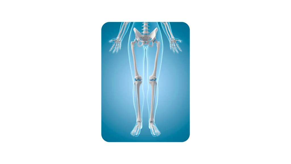
LOWER LIMB BONES
- Phalanges in the fingers.
- The pelvis (ischium, ilium and pubis)
- The femur in the thigh. It is the longest and most powerful bone in the entire skeleton.
- The kneecap in the knee.
- The tibia and fibula, in the leg.
- The tarsus, formed by 7 small bones of the heel.
- The metatarsus in the foot.
Download this lesson as a PDF so you can study offline.