MUSCLES
Muscles are the organs that generate movement in people, THEY ARE THE ENGINES OF HUMAN MOVEMENT. They generate movement by contracting. In the human body the muscles are associated with the skeleton, being responsible of his movement.
They are the active structures of the musculoskeletal system, due to their ability to contract and shorten its length. The 435 muscles of the body represent approximately the 32% of body weight in women and 36% in men. As it contracts, it shortens and the attached bone or structure is pulled. Finished the work, it recovers its position of rest. Globally they have three FUNCTIONS:
- Dynamic, thanks to its contraction the movements are executed.
- Static or postural maintenance by acting on the skeletal pieces, keeping them in a certain position, they carry it out by muscle tension or tone, which is an intrinsic property of muscles. Active ligaments to act as ligaments in the joints stabilizing them and limiting or stopping their movements.
TYPES OF MUSCLES
All the muscles of our body can be classified according to three types of muscle:
Skeletal muscles, striated or voluntary (they are part of the locomotor system, along with the bones and joints):
Characterized by its greater contractility than other cells, excitability, capacity to receive and respond to a stimulus and extensibility-elasticity, capacity from being stretched and returning to its original state after being stretched or contracted.
There are 215 pairs of muscles in the human body. It represents 40% of the weight body in a normal subject. Among its basic functions are: Movement (displacement and carrying out a physical work) and communication
Posture maintenance. Maintenance of the stability of the joints. Heat production.
Smooth, white, involuntary or non-striated muscles:
Ap. respiratory, digestive fundamentally.
Cardiac muscle:
Involuntary. This is the muscle responsible for pumping the blood through the circulatory system by contractions. The myocardium forms the heart walls and is not and is not controlled at will.
MUSCLE COMPONENTS
Connective tissue envelopes:
- Perimysium: Surrounds the muscle.
- Epimysium: Wraps the fascicles.
- Endomysium: Wraps the fibers.
MACROSCOPIC PARTS OF MUSCLE
All muscles have an intermediate portion that we call the body or belly and two ends by which they join the skeletal structures.
From the point of view of muscle biomechanics, muscle insertions are typify in origin and insertion:
Muscular origin or head is the end that during contraction remains fixed and that usually coincides with the end closest to the sagittal plane.
Insertion is the end that moves during contraction and is usually the most distal end.
TENDONS
The tendons are the part of the muscles that does not contract, that is, they have a constant length. They are characterized by being very resistant and flexible, which allows them to conform to bone surfaces or angle under pulleys to change address.
The strength of a tendon is similar to the bones and half that of steel. They are made up of voluminous collagen fibers, in the same direction, which are grouped in fascicles, separated by longitudinal planes of loose connective vascularized tissue.
They have a whitish color, in the form of a cord or smooth ribbon, with round profiles or oval. They are attached to the bone by fibers that attach to the periosteum and penetrate the bone tissue (perforating fibers), which explains bone detachments in certain injuries. Most of them are surrounded by synovial sheaths that protect them.
MICROSCOPIC PARTS OF MUSCLE
Muscle fiber means the structural and functional unit of the muscle, with a imperceptible thickness it size can be between 1 to 50 mm. It is a multinucleate cell (about 100 nuclei), with the ability to become excited and respond to stimuli with a contraction.
The muscle fiber is composed of myofibrils, it is the contractile element of the same, a muscle fiber of 1 cm can contain 8000 myofibrils, constituting the contractile element. Myofibrils are formed by the lengthwise succession of smaller functional units of contraction known as sarcomeres.
Each sarcomere is delimited by “Z” lines, protein frameworks located in the ends that serve to give it stability. Crimped in them and heading toward the center of the sarcomere are thin filaments known as actin filaments.
Occupying the center of the sarcomere, extending towards the ends, one can see a thicker filaments, whose main component is myosin.
CLASSIFICATION OF MUSCLES
Muscles can also be classified according to different criteria as shows in the table:
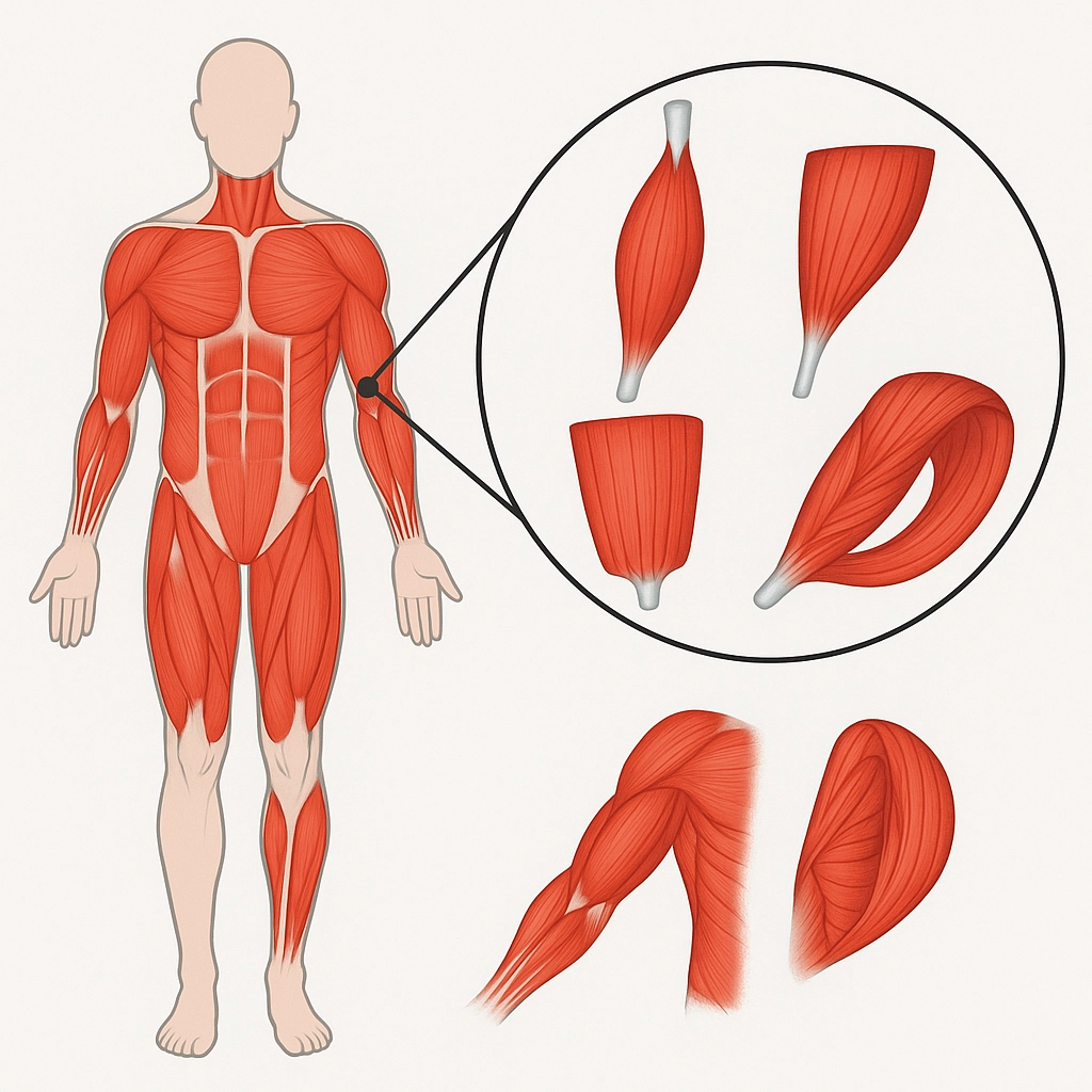
TYPES OF CLASSIFICATION OF STRIATED MUSCLE:
- Due to its relationship with the fusiform tendon: unipenniformes, bipenniformes, multipenniformes.
- By its morphology: Wide, flat and short
- By its type of insertion: Fleshy, aponeurotic and tendinous
- By its number of wombs, origins or insertions: Monogastric, digastric and polygastric, biceps, triceps and quadriceps bicaudal, tricaudal or quadricaudal
- By its movement function: Flexors, extensors, etc.
- Action: agonist, antagonist, fixative or synergist
- By their number of joints: monoarticular, biarticular or polyarticular
But the most useful classification is the one based on the dynamism and behavior in the muscle movement. Thus we distinguish:
Tonic muscles:
They are responsible for maintaining the shape of the body, that is to say that without them we could not stand, we would fall like a meccano. These muscles have a tendency to stiffen, shorten and have contractures muscular.
Phasic muscles:
It can be said that they are in charge of movement, they have tendency (if not exercised) to hypotonia and atrophy.
POSTURAL MUSCLES (TONIC)
Tend to shorten
Triceps Suralis
Iliacus
Psoas
Rectus Femoris
Hamstring
Thigh adductors
Quadratus Lumbar
Deep Extensors
Back trapezius, descending part
Pectoralis Major
Brachial biceps
PHASE MUSCLES
Tend to weaken
Gluteus Maximus
Gluteus medius and minimus
Obliques of the abdomen
Inferior fixators of the scapula (Trapezius, p. ascending, horiz.)
Rhomboids
Triceps brachii
You have to stretch them
You have to train them
MUSCLES OF THE ORGANISM
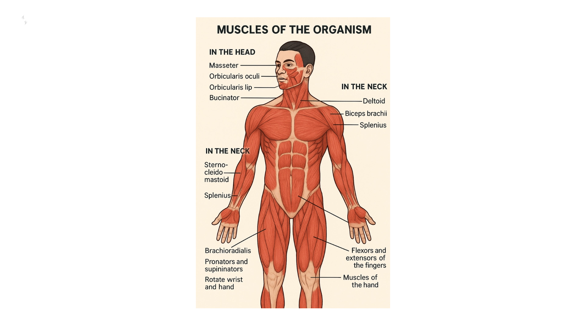
IN THE HEAD
- The ones we use to chew, called masseters.
- The muscle that allows the movement of the lips when we speak: Orbicularis lip.
- Those that allow the eyelids to open or close: Orbicularis oculi.
- The ones we use to blow or whistle, called Bucinators.
IN THE NECK
- The ones we use to bend the head to the sides or to make it turn: Sterno-cleido-mastoid.
- The ones we use to move it back: Splenius.
- The Sternohyoid, which with its contraction lowers the hyoid bone.
IN THE ARMS
- The deltoid that forms the shoulder.
- The biceps brachii which flexes the forearm on the upper arm.
- The triceps brachii which extends the forearm.
- The pronators and supinators rotate the wrist and hand.
- Forearm: Brachiradial.
- Flexors and extensors of the fingers.
- Muscles of the hand.
IN THE LOWER LIMBS
- The buttocks that form the buttocks.
- The sartorius we use to cross one leg over the other. (the longest one of the body)
- The iliac psoas.
- Hamstrings: The biceps femoris is behind, bends the leg at the knee.
- The quadriceps is in front, extend leg.
- The tibialis anterior.
- The twins are the ones we use to walk, they form the calf, they end in the so-called Achilles tendon.
- The soleus.
- Flexors and extensors of the fingers.
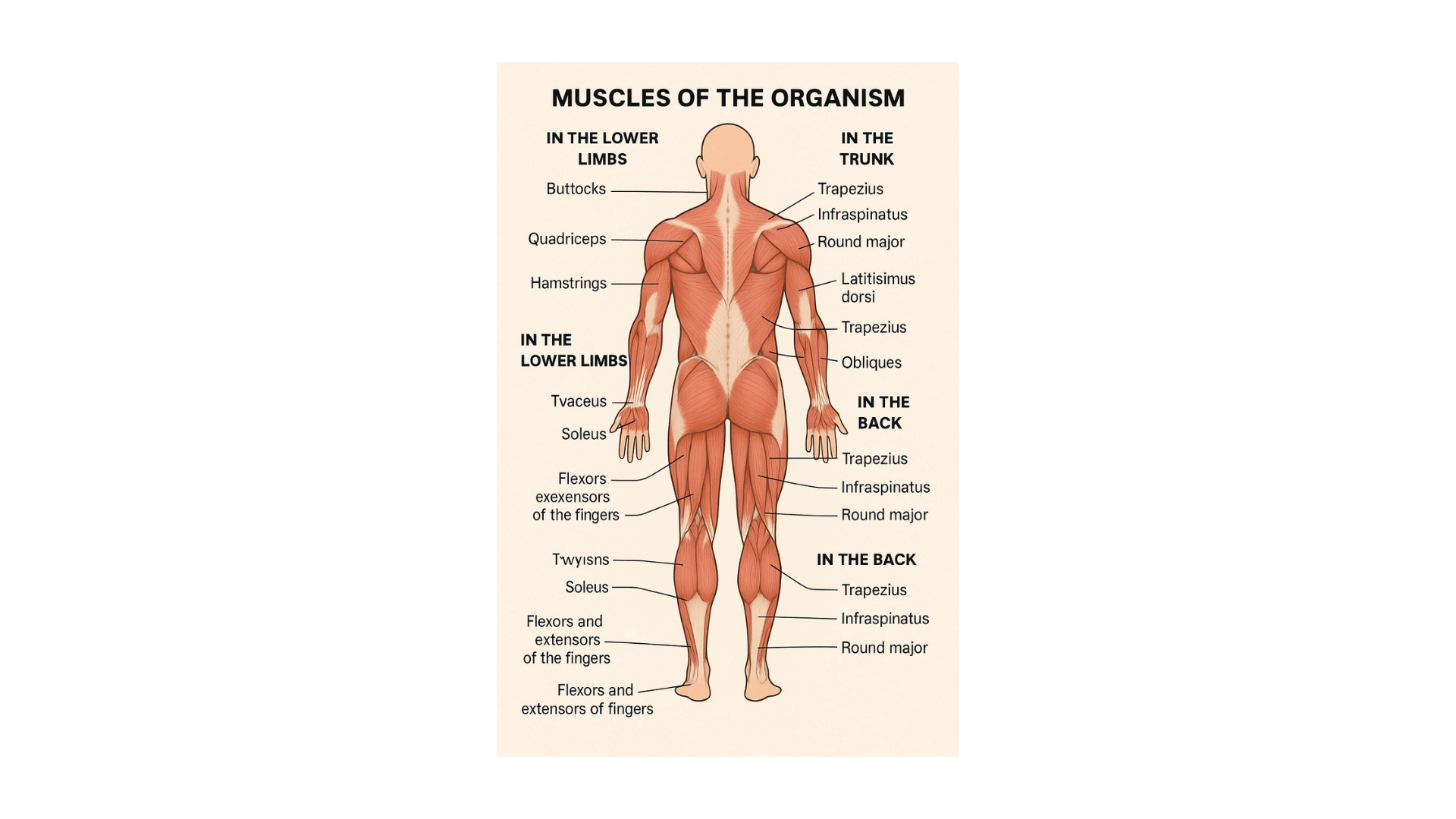
IN THE TRUNK
- Those used in breathing: Intercostals, Serratos, in the form of a saw, the diaphragm that separates the chest from the abdomen.
- The pectorals, to move the arm forward and the dorsal ones, which move the arm back.
- The trapezius, which raise the shoulder and keep the head vertical.
- The obliques of the abdomen (external and internal).
- The rectus abdominis.
IN THE BACK
- Trapezius (on both sides of the spine)
- Infraspinatus (external rotator of the shoulder)
- Round major.
- Latissimus dorsi (largest, widest and strongest of the entire region) acts as an extender and shoulder approdximator.
Download this lesson as a PDF so you can study offline.