DEFINITION OF ARTICULATIONS
A set of elements by which the bones are joined together, allowing the movement of different body segments.
Components of a Joint
- Bone
- Cartilage: Covers the bony surfaces
- Ligaments: Reinforce the joint capsule. Typically four—anterior, posterior, and two lateral
- Capsule: Surrounds the bony surfaces and keeps them in contact
- Synovial Membrane: Thin, transparent; secretes synovial fluid
- Synovial Fluid: Nourishes joint cartilage and lubricates articular surfaces
Additional Joint Structures
- Articular Menisci or Rims: Fibrocartilages found in knee and temporomandibular joints. Essential for knee stability; function as shock absorbers
- Intra-articular Ligaments: Present in the knee (cruciate ligaments) and hip (round ligament)
TYPES OF JOINTS
Classified by anatomical shape and degree of mobility
Mobile Joints (Diarthrosis)
- Space between joint surfaces
- Surfaces covered by articular cartilage
- Joined by joint capsule and ligaments
- Inner capsule lined by synovial membrane producing synovial fluid
- Allows wide range of motion
- Examples: Knee, shoulder, hip joints
Semi-Mobile Joints (Amphiarthrosis)
- Limited mobility
- Bones articulated solidly with a small intermediate space
- Space occupied by intervertebral disc enabling limited extension movements
- Function more as suspension mechanisms than for movement
- Examples: Intervertebral joints, pubic symphysis, sacroiliac joints
Immobile Joints (Synarthrosis)
- Bones joined with no spaces between them
- Joined by suture (toothed, scaly, or harmonic) or interlocking protrusions/concavities
- No mobility
- Examples: Joints of skull vault, face, chondrocostal joints
BASIC STRUCTURE OF SYNOVIAL JOINTS
Formed of components enabling adaptation to extensive movements
Articular Cartilage
- Hyaline cartilage covering articular bone surfaces
- Protects against wear from friction
- Thicker in lower extremities or high-pressure areas
- Nutrient diffusion from synovial fluid and subchondral bone (seen in young individuals)
Synovial Membrane
- Thin and transparent
- Lines the inner joint capsule and bony surfaces
- Forms folds and sacs for movement adaptability
Synovial Fluid
- Transparent, pale yellow liquid
- Non-coagulating
- Plasma dialysate
Menisci
- Located in joints with convex articular surfaces
- Firmly attached to joint capsule and adjacent bones
- Avascular and non-innervated
- Enhance joint stability and congruence
Joint Capsule
- Tough, slightly elastic fibrous tissue sleeve
- Continuation of periosteum
- Surrounds bones and joint structures
- Varies per joint; can be highly visible or barely discernible
- Well-vascularized and innervated

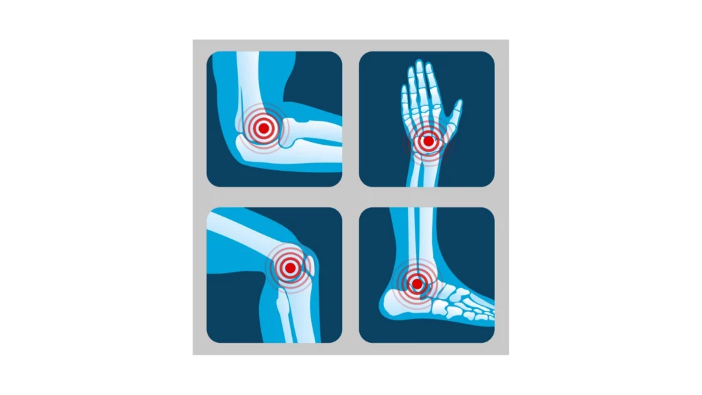
JOINTS OF THE HUMAN BODY
Knee Joint
- Formed by: Femur, Tibia, Fibula (ball joint)
Ankle Joint
- Formed by: Tibia, Fibula, Talus
Wrist Joint
- Formed by: Ulna, Radius, Carpals
Shoulder Joint
- Formed by: Humerus, Scapula (shoulder blade), Clavicle
Hip Joint
- Formed by two joints
- Sacro-iliac: Sacral and Iliac bones
- Coxo-femoral: Coxal and Femur bones
Download this lesson as a PDF so you can study offline.