RESPIRATORY FUNCTION AND RESPIRATORY SYSTEM
- The respiratory system provides the body with oxygen and eliminates carbon dioxide produced in the cells.
- This process is involuntary and automatic: air containing oxygen is drawn in (inspiration), and waste gases including CO₂ are expelled (expiration)
ANATOMY OF THE RESPIRATORY SYSTEM
Divisions of the Respiratory System:
- Airways
- Lungs
AIRWAYS
Upper Airways:
- Mouth and Nostrils:
- Air enters through the nostrils
- Nostrils are lined with highly vascularized mucosa and nasal turbinates
- Functions: warm, moisten air and prevent coarse particles from entering the respiratory system
- Pharynx:
- Tube-shaped structure connecting mouth and nostrils to the larynx
- Larynx:
- Entrance to the respiratory tract
- Contains vocal cords (organ of phonation)
- The epiglottis, a cartilage, protects the larynx during swallowing
Transition Pathway:
- Trachea:
- Flattened tube running from the neck to the thorax
- Composed of ~20 incomplete cartilaginous rings joined by a fibro-elastic membrane
- Divides into two branches: the bronchi
Lower Airways:
- Bronchi and Bronchioles:
- Two main bronchi: right bronchus descends vertically to the right lung, left bronchus goes transversely to the left lung
- Bronchi subdivide up to 25 times, forming the bronchial or respiratory tree
- Bronchioles (diameter <1 mm) end in alveolar clusters
- Alveoli:
- Tiny hollow sacs with thin walls
- Site of gas exchange in the lungs
- Over 300 million alveoli in adult lungs
- Surface area: 70–100 m²
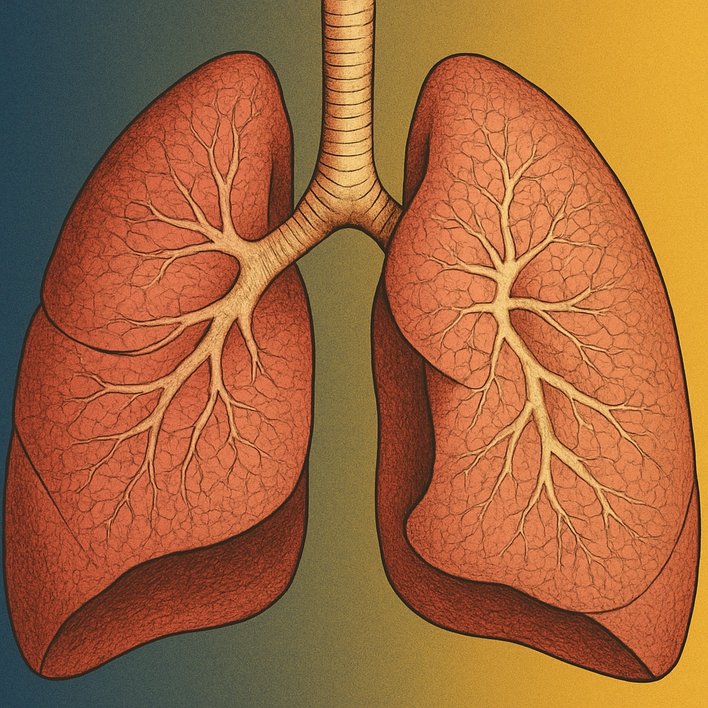
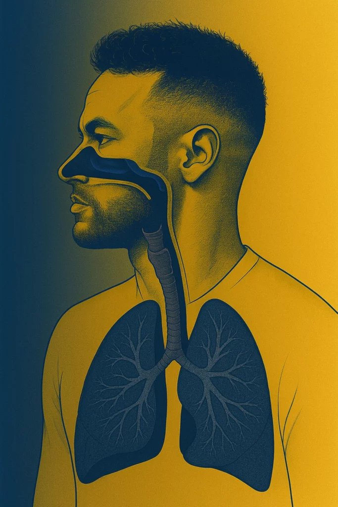
THE LUNGS
- Two spongy reddish masses for gaseous exchange between blood and air
- Shape: Conical with apex at the base of the neck and base resting on diaphragm
- Mediastinum: Space between lungs containing heart, great vessels, and esophagus
- Structure: Each lung contains bronchial branches ending in pulmonary alveoli
THE BREATHING
Definition: Physiological act of oxygen intake and CO₂ release
Phases of Breathing:
- Exchange in the lungs
- Transport of gases
- Respiration in cells and tissues
Exchange in the Lungs:
- Inspiration:
- Rib cage volume increases
- Lungs swell
- Diaphragm and external intercostal muscles raise the ribs
- Expiration:
- Rib cage volume decreases
- Lungs compress
- Upper airways act as air conditioning system
- Breathing rate: 14–16 times/minute
- Air intake per breath: ½ liter
- Lung capacity: ~5 liters
THE TRANSPORT OF GASES
- Oxygen:
- Taken up in alveoli
- Carried by red blood cells to heart
- Distributed via arteries to body cells
- Carbon Dioxide:
- Carried by red blood cells and plasma
- Transported to heart via venae cavae
- Expelled by lungs
RESPIRATION IN CELLS AND TISSUES
- Cells take oxygen from blood to burn absorbed food
- Produces:
- Energy
- Heat (maintains body temperature at ~37°C)
RESPIRATORY ADAPTATIONS DURING EXERCISE
- Increased muscle metabolism requires enhanced:
- Ventilation
- Circulation
- Gas diffusion
Phases of Ventilation During Exercise:
- Rapid rise at exercise start
- Slower sustained ascent
- Post-exercise decrease (repayment of oxygen debt)
- Increased pulmonary blood flow enhances gas exchange and oxygen availability to muscles
- Diffusing capacity:
- Resting: 23 ml/min
- Max exercise: 64 ml/min
LONG-TERM RESPIRATORY SYSTEM ADAPTATIONS
- Enhanced respiratory efficiency and economy
- Increased alveolar respiratory surface
- Improved alveolar-capillary diffusing capacity
- Enlarged pulmonary capillary network
Download this lesson as a PDF so you can study offline.